Dopaminergic pathway on the brain Biology Diagrams
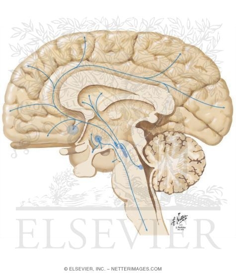
Dopaminergic pathway on the brain Biology Diagrams Fig. 1. (A) Cartoon representation of a sagittal section through the human brain showing the major projections of dopamine neurons from the cell bodies in the ventral tegmental area and substantia nigra (VTA/SN) to terminal field areas in the orbitofrontal cortex (OFC), nucleus accumbens (N. acc.), and dorsal striatum.(B) Cartoon representation of a typical dopaminergic synapse in the brain.

Midbrain dopaminergic (DA) neurons are located in three major nuclei, including the substantia nigra pars compacta (SNpc; A9 group), the ventral tegmental area (VTA; A10 group), and the retrorubral field (A8 group) (Figure 1A).DA neurons in SNpc project to dorsal striatum via the nigrostriatal pathway, and regulate voluntary movement control as part of the basal ganglia circuitry. The great majority of dopaminergic neurons in the brain (in human 300,000-400,000 cells) is organized in three nuclei, the substantia nigra pars compacta, the ventral tegmental area and the arcuate nucleus. The tuberoinfundibular intermediate-length dopaminergic system controls prolactin release from the anterior pituitary and its

Dopaminergic Systems in the Brain and Pituitary Biology Diagrams
Dopamine (dopaminergic) system plays an important role in central neural system, taking part in regulating motor control, executive functions, motivation, reinforcement, reward, sleep, feeding, attention, cognitive functions, olfaction, vision, hormonal regulation, and outside of central neural system in sympathetic regulation and penile erection.

D1-like dopamine receptor expression and its functions. The D1-like receptor family consists of 2 types of GPCRs that include the D1 and D5 receptors, with a higher density in the striatum or caudo-putamen, nucleus accumbens (NAcc), SN pars reticulata (SNr), and olfactory bulb (OB). 31,32 A moderate expression of D1 receptors has been reported in the entopeduncular nucleus, cerebral aqueduct
Dopaminergic pathways Biology Diagrams
Basic dopamine anatomy. The dopaminergic innervation of the forebrain of mammals is constituted by a small number of highly collateralized neurons (~15,000 - 20,000 on each side of the rat brain) residing in the ventral mesencephalon (Fallon and Loughlin, 1995; Lindvall et al, 1984; Williams and Goldman-Rakic, 1998). Three major divisions of dopaminergic pathways innervate the forebrain and It is now 20 years since Swedish scientists described the existence of the nigrostriatal, mesolimbic, and tuberoinfundibular dopaminergic (DA) neurons in the rat brain [4, 8, 13, 17, 24, 50]. Since then new types of DA neuronal systems in the brain have been mapped The main dopaminergic pathways of the human brain. Dopaminergic pathways (dopamine pathways, dopaminergic projections) in the human brain are involved in both physiological and behavioral processes including movement, cognition, executive functions, reward, motivation, and neuroendocrine control. [1] Each pathway is a set of projection neurons, consisting of individual dopaminergic neurons.
